|
||||||||||
|
Advertisement |
||||||||||
|
Google Ad space finances and sponsors ENT USAsm Websites. ENT USAsm, Cumberland Otolaryngology or Dr Kevin Kavanagh, MD do not endorse, recommend, referrer to or are responsible for the Advertisements or for the content or claims made in the Advertisements. |
|||||||||
|
|


 The picture on the right shows a retracted right eardrum. The eardrum has increased vascularity and there may be an infectious component to this effusion.
The pars flassida (the portion of the eardrum above the Lateral process of the Malleus) is retracted and starting to from an attic retraction pocket.
 The picture on the right shows a retracted left eardrum and an attic retraction pocket with scutal erosion (arrow) (distruction of the ear canal bone at the top of the eardrum.)
The eardrum has increased vascularity and there may be an infectious component to this effusion.
 The picture on the right shows a retracted eardrum with air bubbles behind the eardrum.
The picture is displayed with a high contract technique to accentuated the fluid behind the eardrum.
  The picture on the right shows a retracted eardrum with serous fluid and air bubbles behind the eardrum.
 The picture on the right shows a retracted eardrum whose middle ear cavity is filled with serous fluid.
 The picture on the right shows a retracted eardrum. The middle ear cavity has clear serous fluid behind it as demonstrated by a barely visible air fluid level (arrows).
 The picture on the right shows a retracted eardrum with fluid and multiple bubbles in the middle ear. The eardrum is also draped over the long arm of the incus.
 The picture on the right shows a distended eardrum. The patient has just forced air into his middle ear by forced valsalva. Bubbles can be seen behind the upper half of the eardrum and orange serous fluid below this.
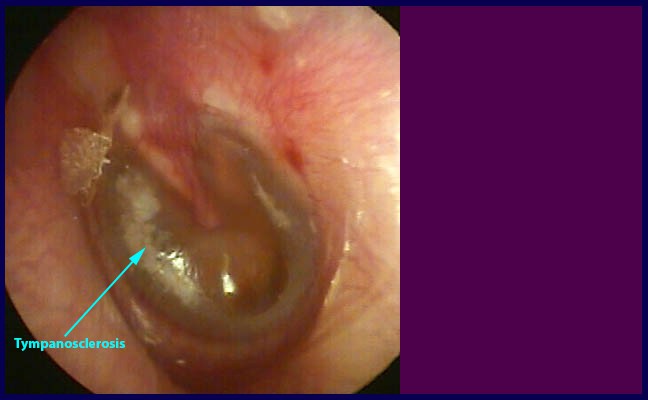
 The picture on the right shows a retracted eardrum. The eardrum has tympanosclerosis and thick glue like fluid in the middle ear.
 The picture on the right shows a retracted eardrum. There is orange fluid in the middle ear and the beginnings of an anterior superior retraction pocket with a very small air bubble.
 The picture on the right shows a retracted eardrum. The eardrum has multiple air fluid levels and air bubbles behind it.
 The picture on the right shows a retracted eardrum.
The eardrum is draped over the promontory and has air bubbles in the middle ear.
 The picture on the right shows a retracted eardrum. There is a large attic retraction pocket with scutal erosion exposure of the neck of the malleus.
 The picture on the right shows a retracted eardrum with orange serous fluid in the middle ear. There is a near normal light reflex and an anterior superior air bubble. The long arm of the incus can also be seen through the eardrum.
 The picture on the right shows a severely retracted eardrum.
The eardrum has mild tympanosclerosis and is forming three retraction pockets. One anteriorly , a second in the posterior-superior quadrant which is starting to erode the long arm of the incus forming a myringoincudopexy, and a third attic retraction pocket with scutal erosion and the beginnings of exposure of the head of the malleus.


