|
|||||||||
|
Advertisement |
|||||||||
|
Google Ad space finances and sponsors ENT USAsm Websites. ENT USAsm, Cumberland Otolaryngology or Dr Kevin Kavanagh, MD do not endorse, recommend, referrer to or are responsible for the Advertisements or for the content or claims made in the Advertisements. |
||||||||
|
|


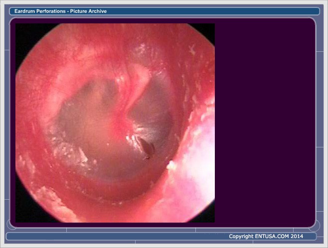 This picture shows a right eardrum with an anterior inferior central perforation. This perforation was caused from barotrauma.
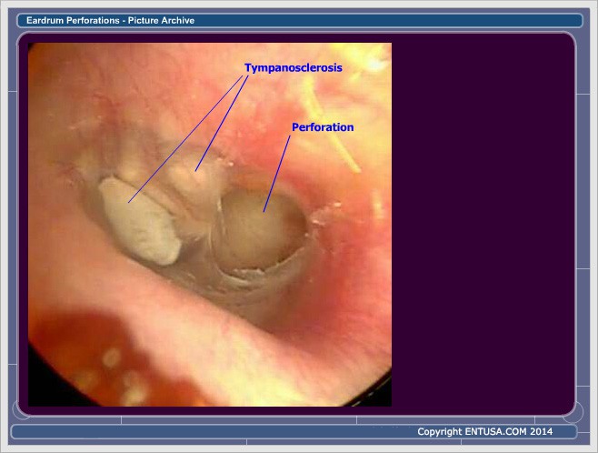 This picture shows a left eardrum with a posterior inferior perforation. Note the two areas of tympanosclerosis.
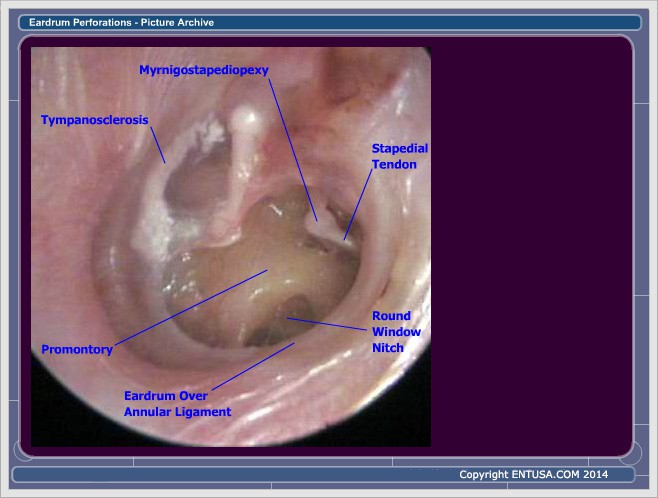 This picture shows a left eardrum with a large perforation. Since the annulus is still present, the perforation is considered to the "central". There also appears to be a tenous connection between the incus and the stapes.
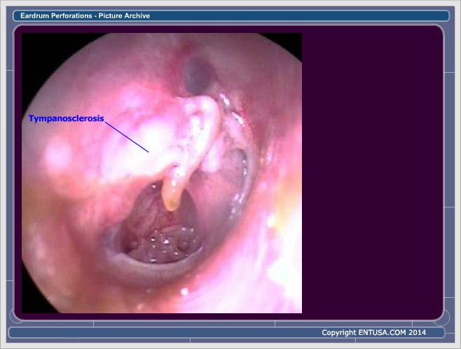 This picture shows a right eardrum with a dry inferior central perforation and a large thick tympanosclerotic plack in the posterior-superior portion of the eardrum.
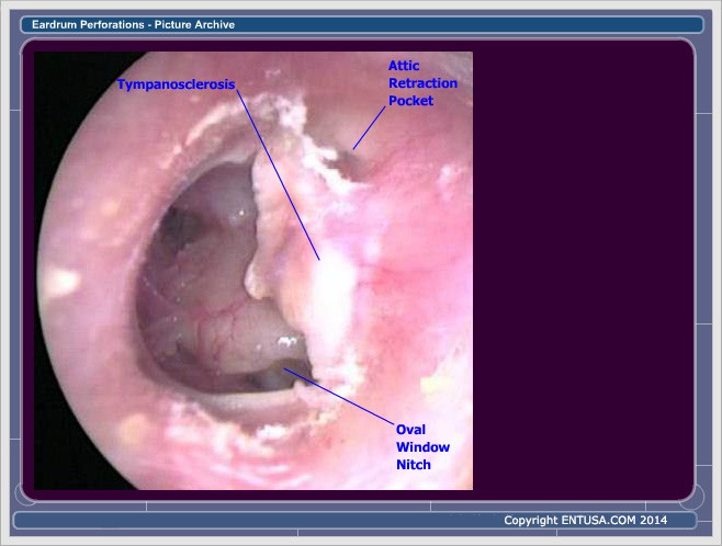 This picture shows a left eardrum with a large perforation involving 80% of the eardrum and a thick tympanosclerotic plack in the posterior-superior portion of the eardrum.
 This picture shows a right eardrum with a posterior central perforation and a tympanosclerotic plack in the anterior portion of the eardrum.
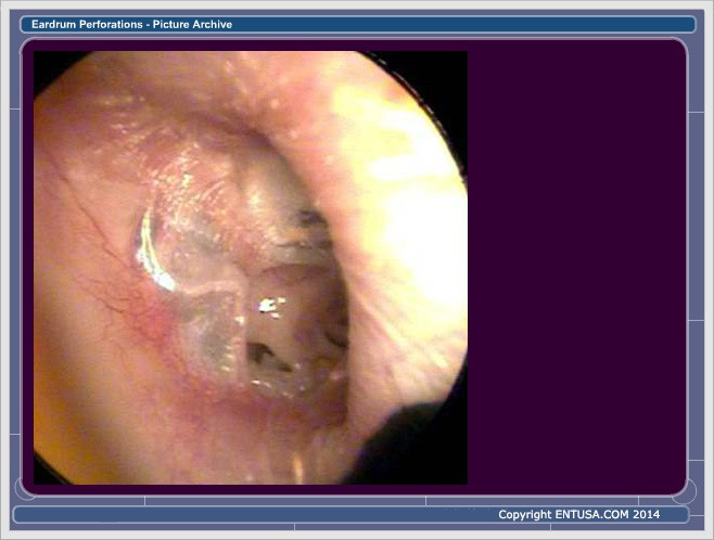 This picture shows a right eardrum with an inferior central perforation and anterior tympanosclerosis.
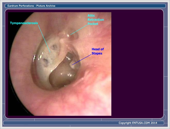 This picture shows a left eardrum with a posterior perforation and anterior tymapnosclerosis. Note the attic retraction pocket and erosion of the arm of the inus with loss of its connection to the head of the stapes.
 This picture shows a right eardrum with a posterior central perforation and anterior tympanosclerosis.
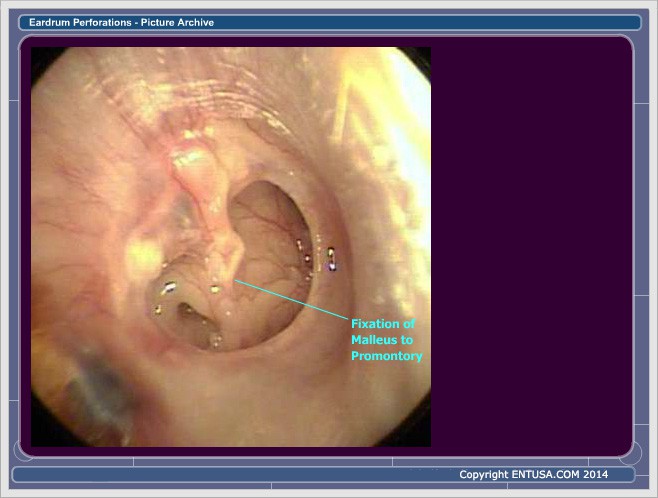 This picture shows a right eardrum with a large inferior central perforation. Note the fixation of the umbo (malleus) to the promontory.
 This picture shows a left eardrum with a large central perforation.
 This picture shows a right eardrum with an posterior marginal perforation. Marginal perforations are at risk for skin growing from the ear canal into the middle ear which can form a cholesteatoma.
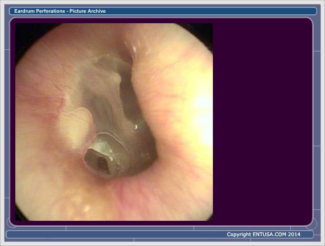 This picture shows a right eardrum with an posterior marginal perforation. Marginal perforations are at risk for skin growing from the ear canal into the middle ear which can form a cholesteatoma.
 This picture shows a right eardrum with a wet anterior superior central perforation. The eardrum has extensive tympanosclerosis and is inflamed next to the posterior rim of the performation.
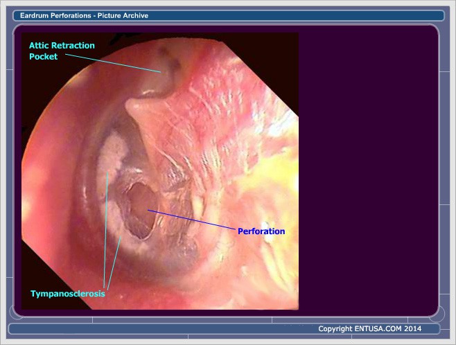 This picture shows a left eardrum with an anterior inferior central perforation, small areas of tympanosclerosis and an attic retraction pocket.


